- Home
- Features Index
-
Features
Features
The Final Intellectual Frontier: Unraveling the Mysteries of the Brain
2024/12/09
The brain, a realm as vast and uncharted as the Earth and the cosmos itself, is often referred to as “humanity’s final intellectual frontier.” Nestled within the halls of Keio University's School of Medicine, the Division of Brain Sciences stands at the forefront of this frontier, part of the Institute for Advanced Medical Research's quest to unravel the mysteries of the mind and pioneer groundbreaking treatments for mental disorders, which are the result of breakdowns in brain function. We sat down with the head of the lab, Prof. Kenji Tanaka, to hear about their recent research achievements, the potential of their work, and the pride they have as researchers who dare to explore the unknown.
Driven by a Desire to Understand the Mechanism of Recovery
"I was just curious about the mind," reveals Prof. Kenji Tanaka when asked about his surprisingly vague reasons for joining the Department of Neuropsychiatry at Keio University School of Medicine. He credits his pivotal shift toward research to a profound encounter with a patient suffering from a mental disorder during his clinical residency.
“I was astonished at how mental disorders could plunge people into such misery. Even more shocking was seeing how dramatically symptoms could improve with medication. Yet, despite reading every textbook I could find, I still couldn't understand why these drugs worked. It was like a black box that ignited a desire in me to unravel the mechanism of recovery.”
For his graduate studies, Prof. Tanaka joined the Department of Physiology, led by Professor Emeritus Keiichi Uemura, a luminary in neurochemistry. He later honed his skills at the Fujita Health University Institute for Comprehensive Medical Science (ICMS) and the National Institute for Physiological Sciences.
"This was back in the early 2000s, when genes responsible for hereditary neurological disorders were being identified one after another. While more and more was being revealed about neurons, the functions of other types of glial cells remained poorly understood and garnered little attention. I began my research driven by the belief that studying them might reveal something new. Prof. Kazuhiro Ikenaka at the National Institute for Physiological Sciences was very supportive and encouraging. He told me that he wasn’t sure if studying glial cells would help in understanding mental disorders, but if I wanted to explore them, I should go ahead.”
It’s been almost 25 years since Prof. Tanaka was, in his own words, a "virtually clueless” medical student, knocking on the door of the Department of Neuropsychiatry. Today, he leads over 30 researchers and medical students at the Division of Brain Sciences in the Institute for Advanced Medical Research.
“Seeing patients get better, being able to do things they couldn't do before— I've spent my career exploring the brain mechanisms behind these dynamic changes. The Division of Brain Sciences in the Institute for Advanced Medical Research is dedicated to understanding how to improve brain function and alleviate brain dysfunction. At the same time, it forced us to grapple with the grand question: What is the human brain?”
The Innovative Technologies Transforming Brain Science Research
Composed of billions of cells, each receiving over 10,000 inputs, the brain's immensely complex structure and function have gradually been elucidated alongside the evolution of technology.
For instance, the introduction of computed tomography (CT) and magnetic resonance imaging (MRI) in hospitals during the 1970s and 80s enabled the visualization of information within the living body and brain. Moreover, the widespread use of green fluorescent protein (GFP) since the 1990s has allowed the observation of cellular activities under a light microscope without compromising cell viability. In 2009, single-cell sequencing emerged, a technique capable of examining gene expression in individual cells. These advancements deepened our understanding of cell classification and states, significantly advancing the elucidation of the brain's structure.
However, Prof. Tanaka notes that there were still challenges in clarifying the brain's function.
“Identifying the causal relationship between specific neural activities and behaviors has been extremely challenging. Even in animal experiments, electrical stimulation spreads too broadly, making it impossible to manipulate only specific neurons. Without a direct method of proof, the relationship between neural activity and behavior was traditionally just speculation based on correlations. For instance, take the striatum—an essential part of the basal ganglia. When examining this part of the brain, which is a group of nuclei in the brain crucial for controlling movement and coordination, researchers were limited to making educated guesses regarding its neural functions. They speculated that within this structure, neurons equipped with dopamine D1 receptors were likely to encourage movement, while those with dopamine D2 receptors were thought to suppress it, yet definitive proof remained elusive.”
But then, a breakthrough came in 2005 with the advent of optogenetics.
“Optogenetics is a technique that allows for the precise manipulation of target cells on a millisecond scale through the toggling of light exposure, which enables the control of cellular activity within the body with high temporal and spatial resolution.For the first time, it became possible to empirically demonstrate the causal relationship between neural activity and behavior, significantly accelerating the elucidation of the function of neural cells.”
In fact, Prof. Tanaka was involved in the early days of this groundbreaking technology.
“I was studying at Columbia University in 2006, working in the lab of Prof. René Hen, where Karl Deisseroth, the developer of optogenetics, would often visit. He'd come into the lab saying, 'I've developed a new technology. Try it out.' Initially, it was a basic tool, only working on cultured cells, and it took several years of trial and error before it could be used in live mice. I'm honored to have been part of the process of refining this excellent technology and making it more widely accessible. Optogenetics has now become an indispensable tool in brain science research, used to manipulate neural cells as well as glial and vascular cells.”
Deciphering the Neural Foundations of Motivation: Promising Advances for Treating Depression
Ambitious large-scale research projects are underway worldwide aiming to fully decipher and create a "map" of the human brain. Following the launch of the United States' BRAIN Initiative in 2013 and Europe's Human Brain Project, Japan began the Brain Mapping by Integrated Neurotechnologies for Disease Studies (Brain/MINDS) project in 2014.
"Despite the tireless efforts of the world's most brilliant researchers, the brain still harbors many unknown territories. I find it incredibly rewarding and am grateful for the opportunity to be one of the people tasked with taking on this challenge.”
One of the recent achievements of Prof. Tanaka and his research team was in the study of motivation.
“In conditions such as depression, dementia, and cerebrovascular disorders, a common symptom is a decrease in motivation. Conversely, with addiction, there is an abnormal increase in motivation. The neural basis for these motivational abnormalities is largely unknown, and there are currently no treatments available for impaired motivation, especially following brain injury.”
Prof. Tanaka’s research team, drawing on cases of motivation decline in brain injuries and neurodegenerative diseases, focused on dopamine receptor type 2-expressing medium spiny neurons (D2-MSNs), which are components of the ventrolateral striatum deep within the brain. They created genetically modified mice in which these D2-MSNs gradually die off, mimicking neurodegenerative conditions. The mice were first trained to understand that pressing a lever would yield a food reward, the assumption being that mice who pressed the lever more frequently exhibited higher motivation. The team discovered that sufficiently motivated mice showed a decline in motivation, with just a 17% cell death in the ventrolateral striatum. Furthermore, by inhibiting D2-MSN function through optogenetics, Prof. Tanaka and his team revealed that D2-MSNs in the ventrolateral striatum are essential for initiating motivated behavior. This discovery of neurons responsible for motivated behavior—essentially the neurons that say “let's do this!”—was first published in Nature Communications in 2017.
In 2019, the research team announced a study focusing on persistence, the ability to sustain motivated behavior. It revealed that during motivated behavior, the activity of neurons in the ventral hippocampus decreases due to serotonin and demonstrated that the inhibition of neuronal activity in the ventral hippocampus is essential for the continuation of motivated behavior.
“We've discovered that the initiation of motivated behavior, the ‘let's do this!’ moment, and the continuation of motivated behavior, the ‘let’s make it to the end’ process, are each controlled by different brain regions and neurons. Additionally, we found that when neurons in the ventromedial striatum are activated, there's an increase in unnecessary actions that inhibit goal-directed behavior, which we are currently studying as ‘fickle’ neurons. Unraveling the neural foundations of motivation, persistence, and fickleness is expected to lead to a better understanding of the mechanisms behind fluctuations in motivation and the development of new treatments as well as to insights into conditions such as adjustment disorders and obsessive-compulsive disorders, which result in a lack of cognitive flexibility.”
Aiming to Shed Light on Human Diseases through the Study of Structural Abnormalities
Recently, there has been a growing interest in a new approach to analyzing brain structure abnormalities through MRI brain images of humans and mice.
“This approach is based on the idea that the differences between healthy individuals and those with mental disorders lie in the function of the brain, which manifests in the structure and shape of the brain. This lets us answer questions like, ‘What is the difference between a brain with a methamphetamine dependence and one without?’ Or, ‘What part of the brain changes when a patient with depression is treated with antidepressants?’ While this is currently limited to laboratory animals, investigations using MRI and electron microscopy have shown that changes occur in the parts of the brain known as the cingulate cortex and ventral pallidum. respectively.”
In 2023, Prof. Tanaka and his team published the discovery of a mechanism—how dyskinesia, a drug-induced movement disorder in Parkinson’s disease, progresses—which was also a breakthrough for detecting changes in the brain's structure.
Patients with Parkinson’s disease who take the medication Levodopa over a long period can develop a difficult-to-treat side effect known as Levodopa-induced dyskinesia (LID), characterized by extra, involuntary movement of the body and limbs. Similarly, patients with mental disorders may develop tardive dyskinesia (TD), characterized by repetitive and involuntary movements such as grimacing, as a result of the long-term use of antipsychotic drugs.
“Despite the use of different drugs to treat different diseases, the emergence of similar side effects led us to hypothesize that LID and TD might share a common underlying mechanism.”
Upon creating LID and TD model mice and searching for common structural abnormalities in the brain, the team discovered that in both conditions, there was an enlargement of the axon terminals of striatal neurons in the globus pallidus. Subsequently, they also identified that an increase in the expression of the vesicular gamma-aminobutyric acid transporter (VGAT) in striatal neurons was a contributing factor to the development of dyskinesia, and furthermore, that fluctuations in brain dopamine levels lead to the overexpression of VGAT.
“This research led to the identification of genes and the elucidation of their functions, thanks to our observation of the structural changes in the form of cell enlargement in the basal ganglia. We anticipate that similar approaches, leveraging accumulated human brain data, will further advance our understanding of human diseases.”

Embracing the Grind: Persistence as a Researcher
Alongside his research, Prof. Tanaka dedicates one day a week to seeing patients in the neuropsychiatry outpatient clinic.
“Researchers tend to be drawn to problems with potential solutions, but when I’m face-to-face with a patient, I’m reminded of my commitment to research that aims to solve truly pressing problems.”
Prof. Tanaka shared what he thinks makes someone well-suited for neuroscience research.
“Well, this isn't limited to the field of neuroscience, but anyone who is set on the idea that things must go a certain way might not be suited for research.While it’s enjoyable when an experiment unfolds according to your hypothesis, it’s crucial to derive joy in the trial and error when it doesn't. You have to be open to any result, carefully interpret it, and be willing to repeat an experiment any number of times. That’s the attitude a researcher needs to have.”
Isn’t it common to encounter challenging situations that seem impossible to overcome, even through repeated trial and error?
“Of course, there are many times when things don't go as planned, or another researcher beats you to the punch, but I don't worry about those things.Only those who are actively working with their hands and collecting data gain something in the process. Besides, there are times when a result that seemed like a failure because it didn't align with your hypothesis later turns out to be significant, making you realize that it was actually an important finding after all.”
And that glial cell research that Prof. Tanaka began on a hunch in his younger days? He says that research has advanced both domestically and internationally, revealing many important functions of glial cells, but even after more than two decades, they are still far from fully understanding their role in diseases. Nevertheless, in 2021, Prof. Tanaka and his team discovered that a specific molecule expressed in oligodendrocytes, a type of glial cell, can control age-dependent neural plasticity. This finding is expected to contribute to improving brain functions that have diminished due to aging and other factors, and research is steadily progressing.
Challenging But Rewarding: Finding Your Calling
Since being re-established 12 years ago, the Division of Brain Sciences in the Institute for Advanced Medical Research has seen its diverse team of researchers achieve an equally diverse variety of research outcomes. The lab operates under three mottos long championed by Prof. Tanaka: “Beauty is Truth,” “Enjoy the Research,” and “Learn from Your Collaborators.”
“‘Beauty is Truth’ is something I inherited from Prof. Kiyoshi Hama, an Professor Emeritus at the National Institute for Physiological Sciences. The arrangement and shape of cells viewed under a microscope are truly exquisite and beautiful. Interestingly, when we modify genes or manipulate cellular activity with optogenetics, this beautiful structure can sometimes be disrupted. There's a reason behind beauty, and within that beauty lies truth. This sentiment is something that I think resonates with many researchers.”
The second motto, “Enjoy the Research,” he learned from another late mentor, Prof. Kazuhiro Ikenaka.
“Prof. Ikenaka truly embraced his research with a sense of freedom and joy, which I credit for my own perseverance in the field. Regardless of industry or profession, people who enjoy their work seem to be happier, don’t they? I want the next generation to be able to fully immerse themselves in medicine and healthcare with that same sense of joy. That’s really what it comes down to. Of course, along with that joy, there are also challenges and hardships, but the key is to pursuit and see things through.”
The third motto, “Learn from Your Collaborators,” embodies the message that collaborative research should be conducted not for personal gain but to exchange and elevate each other’s skills and knowledge. This is in line with the Keio University principle of hangaku-hankyo—learning while teaching, teaching while learning—in the relationship between teachers and students.
Prof. Tanaka was also kind enough to share a message for the next generation of students aspiring to a career in medicine and healthcare.
“I often hear people say they don't know what they're suited for, and I tell those people to just give things a shot—try a little bit of everything. Of course, if you are interested in neuroscience research, you are more than welcome here. In any case, I think students need to spend their time exploring and finding themselves. I hope you find a path that truly brings you joy and that you give it your all.”
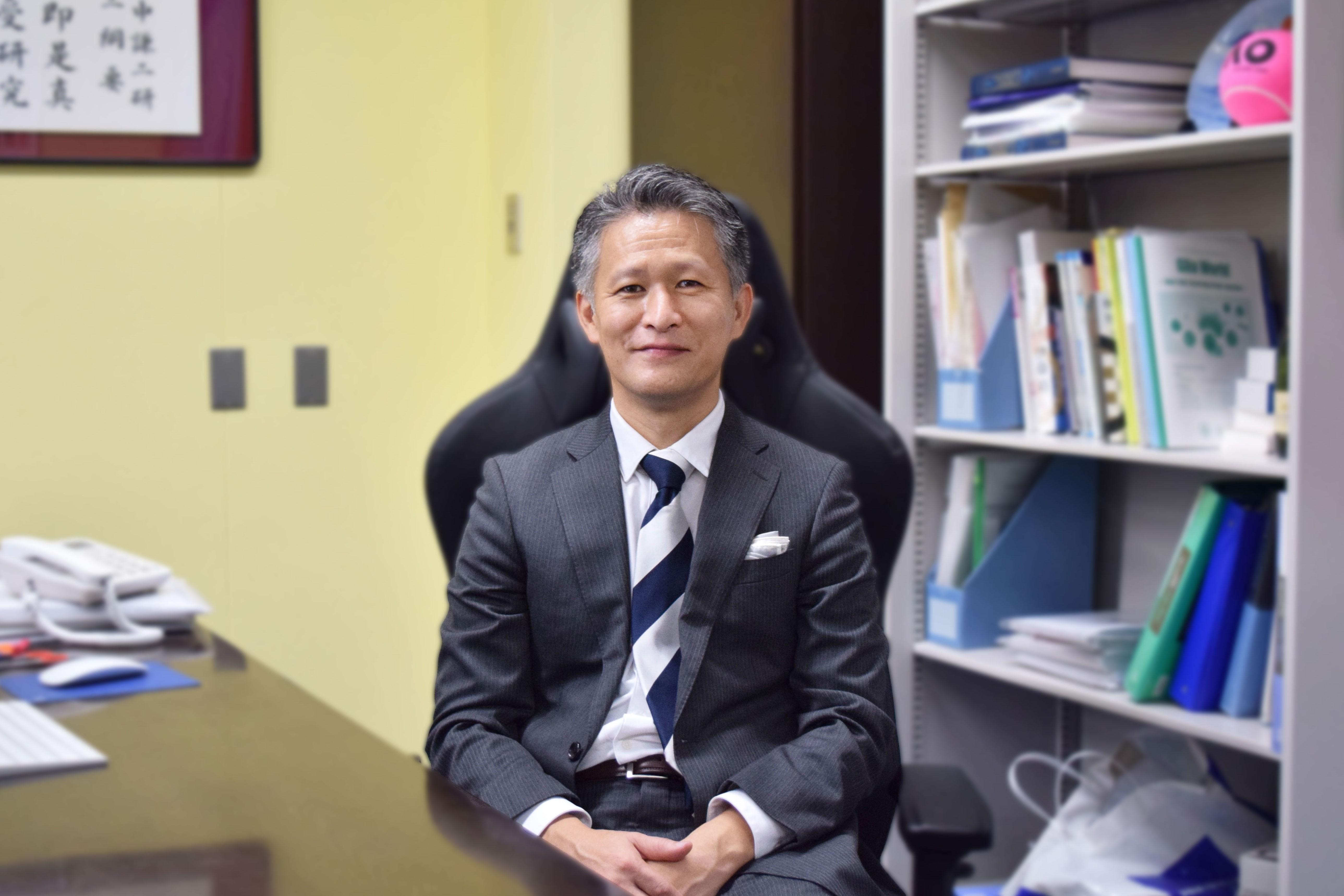
Kenji Tanaka
Kenji Tanaka graduated from the Keio University School of Medicine in 1997 and completed his doctoral program at the Keio University Graduate School of Medicine in 2003.After serving as a postdoctoral fellow at the National Institute for Physiological Sciences and then as a postdoctoral research scientist at Columbia University, he returned to the National Institute for Physiological Sciences, where he became an assistant professor in 2008.He held positions as a Project Associate Professor and Associate Professor in Neuropsychiatry at the Keio University School of Medicine before taking on his current position in 2021.Prof. Tanaka has received numerous awards, including the NARSAD Young Investigator Award, the Sanshikai Young Investigator Award, the Japanese Society for Neurochemistry’s Distinguished Investigator Award, the Tatsuji Nomura Award, and the Kitasato Award.