HOME > Program Members > Yasuyuki Ohnishi

Yasuyuki Ohnishi

General Manager, Central Institute for Experimental Animals
Yasuyuki Ohnishi, DVM, PhD
kiyasho@ciea.or.jp
http://www.ciea.or.jp
Theme
Imaging analysis of the central nervous system of small animals
Our role and primary goal in this program is to develop an optimal imaging strategy for morphological and functional analysis of the central nervous system of small animals. We are conducting this research in collaboration with the Central Institute for Experimental Animals where a 7T MRI unit has been in operation since 2004. So far, we have established an MRI protocol for the assessment of a spinal contusion model. We are currently working on imaging analysis of the normal central nervous system of common marmosets and other small animals.
Research activities
- Diffusion Tensor Neuronal Fiber Tractography and Manganese-enhanced MR Imaging of Primate Visual Pathway in the Common Marmoset
- Functional MRI study of neurovascular coupling of rat
- Brain MR atlas of common marmoset
- MRI study of the spinal contusive model of the common marmoset
- Diffusion tensor tractography of the spinal contusive model of the common marmoset
- q-space MRI of the spinal contusive model of the common marmoset
- Diffusion tensor tractography of the peripheral nerve contusive model of the rat
[Collaborating Researchers]
Masayuki Yamada (School of Health Science, Fujita Health University)
Kenji Kawai (Pathology Research Department, Central Institute for Experimental Animals)
Akio Iwanami, Junichi Yamane, Nobuyuki Fujita, Kanehiro Fujiyoshi, Takehiko Takagi (Department of Orthopaedic Surgery, Keio University)
Miyasaka Naoyuki (Department of Comprehensive Reproductive Medicine, Tokyo Medecal Dental University)
Yoshitaka Masutani (Department of Radiology, University of Tokyo)
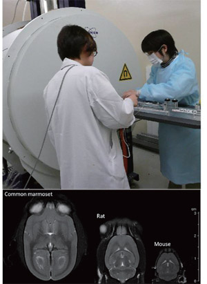
Fig.1
Normal horizontal section (T2 weighted image) of the brain of the small experimental animals (common marmoset, rat, mouse)
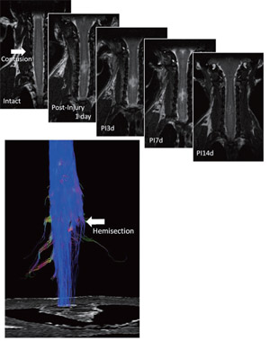
Fig.2
LongitudinalMRI(T2weighted image) in the spinal contusion model of the common marmoset
Diffusion tensor tractography in the spinal hemi-section model of the common marmoset
Selected Paper
- Takagi T, Nakamura M, Yamada M, Hikishima K, Momoshima S, Fujiyoshi K, Shibata S, Okano HJ, Toyama Y, Okano H. Visualization of peripheral nerve degeneration and regeneration: monitoring with diffusion tensor tractography. NeuroImage. 44:884-892, 2009.
- Yamada M, Momoshima S, Masutani Y, Fujiyoshi K, Abe O, Nakamura M, Aoki S, Tamaoki N, Okano H. Diffusion-Tensor Neuronal Fiber Tractography and Manganese-enhanced MR Imaging of Primate Visual Pathway in the Common Marmoset: Preliminary Results. Radiology. 249:855-864, 2008.
- Fujiyoshi K, Yamada M, Nakamura M, Yamane J, Katoh H, Kitamura K, Kawai K, Okada S, Momoshima S, Toyama Y, Okano H. In vivo tracing of neural tracts in the intact and injured spinal cord of marmosets by diffusion tensor tractography. J Neurosci. 27(44):11991-8, 2007.
- Iwanami A, Yamane J, Katoh H, 1. 1. Nakamura M, Momoshima S, Ishii H, Tanioka Y, Tamaoki N, Nomura T, Toyama Y, Okano H. Establishment of graded spinal cord injury model in a nonhuman primate: the common marmoset. J Neurosci Res. 80(2):172-81, 2005.
- Iwanami A, Kaneko S, Nakamura M, Kanemura Y, Mori H, Kobayashi S, Yamasaki M, Momoshima S, Ishii H, Ando K, Tanioka Y, Tamaoki N, Nomura T, Toyama Y, Okano H. Transplantation of human neural stem cells for spinal cord injury in primates. J Neurosci Res. 80(2):182-90, 2005.
Copyright © Keio University. All rights reserved.