- Home
- Features Index
-
Features
Features
Unraveling the Mysteries of the Human Body: The Department of Anatomy Explores the Vascular Network
2023/06/13
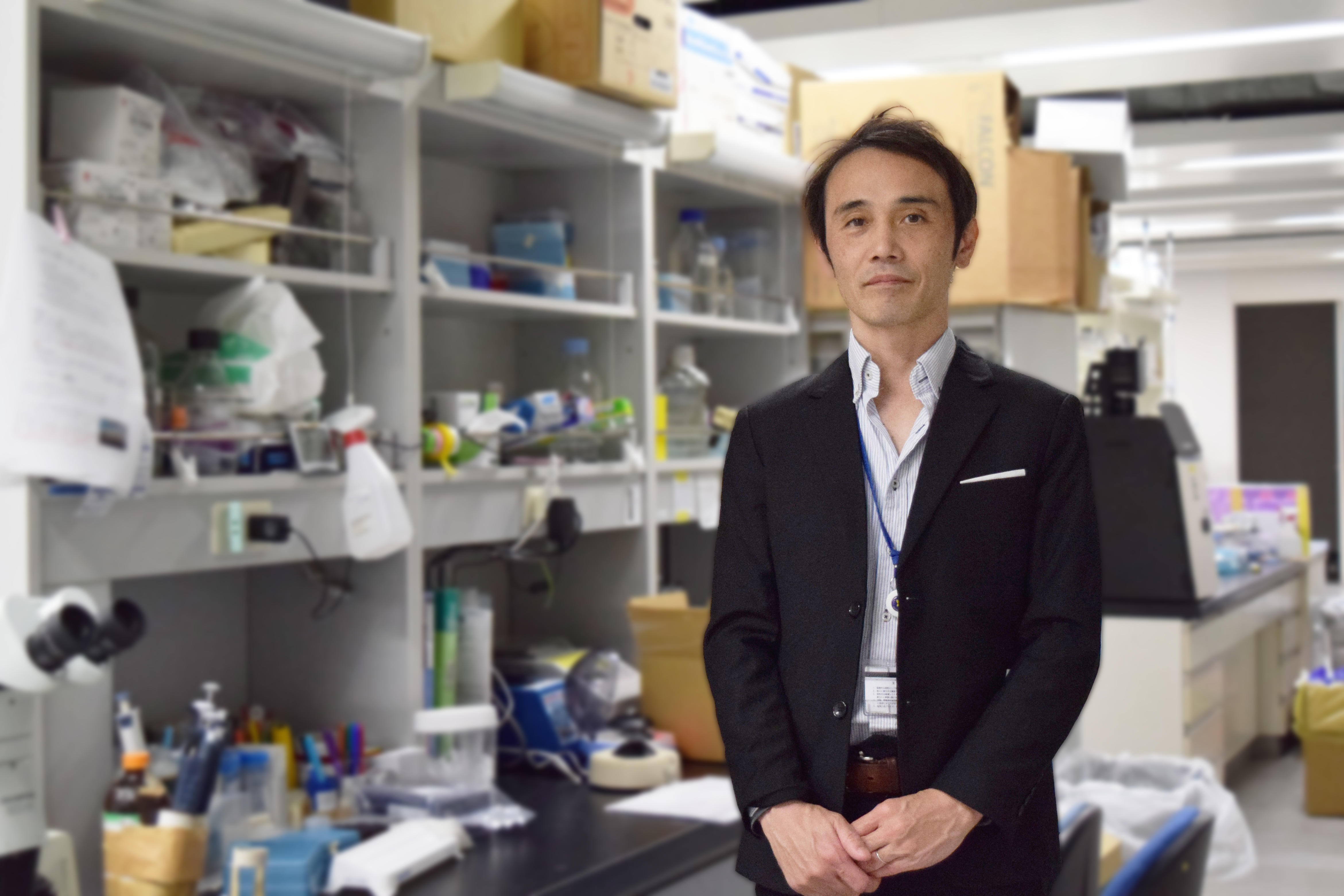
Our bodies are quite literally packed with blood vessels from head to toe, but have you ever wondered how this vascular network is formed? That is the question that Professor Yoshiaki Kubota, head of the Laboratory of Vascular Biology in the Department of Anatomy at the Keio University School of Medicine, is trying to answer. Here, we sit down with Prof. Kubota to discover the pride of a researcher who has come face to face with life’s mysteries and learn what he means when he says, “It's not about doing research that serves a purpose, but rather doing research that you find interesting.” He also shares why he thinks of himself as a bit of an outlier and gives advice to the next generation.
Vascular Network Diagrams Were Made Up This Whole Time?!
“According to one theory, the total length of human blood vessels is 100,000 km, a distance equivalent to two and a half times around the Earth. Don't you think it's peculiar that these blood vessels can run throughout our bodies without getting all tangled up?”
So begins Prof. Kubota, who goes on to say that many of their structures and functions have yet to be fully understood, despite the fact that blood vessels are also thought of as an essential organ for sustaining life and delivering oxygen and nutrients to cells throughout the body.
“The human vascular system is formed in the blink of an eye, taking only about three months from when an egg is fertilized. But what mechanism is behind this process? How do blood vessels split into thin capillaries like branches off the trunk of a tree? And why don't certain areas of the body have blood vessels? Cancer cells use surrounding blood vessels to proliferate, but why are they unable to create normal, healthy blood vessels? These are the kinds of questions we ask in vascular biology, seeking to understand vascular formation at the cellular and molecular level.”
In vascular research, visualizing blood vessels has long been a challenge. Traditional methods of observing cross-sections of blood vessels from tissue slices could only fill in the gaps based on assumptions of what might be happening in between. It wasn’t possible to accurately grasp and observe the vascular network within the living body.
“A turning point came about 15 years ago. Our lab developed high-resolution 3D imaging technology, and research institutions around the world began developing and improving similar technologies to visualize blood vessels. This has made it possible to perform detailed observations of vascular networks. In the field of vascular medicine, there are still organs that we cannot accurately visualize, and sometimes we find blood vessels in states that we cannot explain, so we've seen an expansion in potential applications for this kind of research in recent years.”
Plastic Surgeon Turned Vascular Researcher
Prof. Kubota’s move into vascular research was prompted by his clinical experience in plastic surgery.
"Working in plastic surgery, I often dealt with the treatment of pressure ulcers, or bedsores. Textbooks would always state the importance of increasing the number of blood vessels, so that was the goal with most treatments. But in many cases, a patient's condition didn't improve despite the area appearing red and supposedly rich in blood vessels. I started to think that something was wrong with this approach.”
The conclusion Prof. Kubota reached through his clinical experience and research differed from what his textbooks described.
“I realized that for blood vessels to function properly, it's important to maintain a strict hierarchy— from arteries to capillaries to veins—and ensure that oxygen and nutrients are transported efficiently. In other words, quality is essential. This means that even if you increase the quantity of capillaries, it doesn’t matter unless the blood vessels have the capacity to supply them.”
Doubt and distrust of conventional wisdom became one of the catalysts for Prof. Kubota to pursue a career in research. Encouraged by plastic and reconstructive surgery lecturer (now professor) Kazuo Kishi, he would later go to work in Prof. Toshio Suda's developmental biology laboratory.
“I began my research career investigating the function of molecules essential for blood vessel development in zebrafish. The days I spent working under Prof. Suda were fulfilling, both in terms of ambition and intellectual curiosity and I realized that what I wanted was to pursue a career in research.”
Gradually Unraveling the Mechanism of Vascular Formation
It's been nearly 20 years since Prof. Kubota began researching blood vessels. Today, his laboratory tackles a truly diverse range of research topics.
In 2020, the lab discovered the mechanism that keeps blood and lymphatic systems separate.
“Blood vessels and lymphatic vessels—especially veins and lymphatic vessels—have such similar characteristics and structures that they are almost indistinguishable. Nevertheless, they remain independent systems that never intersect, and the mechanism behind this phenomenon had long remained a mystery,” Prof. Kubota explains.
His research first came about when an acquaintance from Prof. Kubota’s time at the National Institutes of Health in the United States requested an investigation into the Folliculin gene, which can cause Birt-Hogg-Dubé (BHD) syndrome, a condition associated with kidney cancer and other diseases.
“We made thorough observations of Folliculin-deficient mice and found that this deficiency resulted in red blood cells entering lymphatic vessels. We also observed abnormal connections between blood vessels and lymphatic vessels.”
When Folliculin is deficient in vascular endothelial cells, “lymphatic-biased venous endothelial cells” appear in some parts of the blood vessels, causing them to be misidentified as the target for connection to lymphatic vessels. In other words, the lab discovered that Folliculin is responsible for maintaining the segregation of these two networks.
“Finding the mechanism by which these very similar circulatory systems form independent networks attracted attention as a significant discovery in biology.”
In 2022, the lab was the first in the world to establish a high-resolution imaging technique, which they used to reveal the mechanism behind how teeth harden.
“The tooth is the hardest structure in the human body, and cutting one into sections is extremely difficult. It was also considered virtually impossible to observe in detail the blood vessels inside the dental pulp at the center of each tooth. In our study, we successfully prepared sections of mouse teeth and visualized the 3D structure of tooth vasculature by making various improvements to decalcification and immunostaining procedures. We identified a specific group of blood vessel cells important for teeth to harden and were able to uncover the mechanism behind it.”
Research Should Be 90% Fun: Taking Pride in Observational Studies
Prof. Kubota mentions other examples where basic research findings have eventually led to clinical trials. One such example is a study on new blood vessels that play a crucial role in cancer cell proliferation, published in 2012.
Eleven years ago, Prof. Kubota and his team discovered that the ATM (ataxia-telangiectasia mutated) gene, which eliminates reactive oxygen species (ROS) accumulated by ultraviolet and radiation exposure, is actively involved in tumor neovascularization, whereby new blood vessels are formed from existing ones following endothelial cell proliferation and migration. They found that in ATM-deficient mice, excessive accumulation of ROS caused early aging and shrinkage of blood vessels, resulting in poor blood flow to supply oxygen and nutrients to cancer cells, thereby suppressing cancer growth.
“Researchers continue to aim for clinical application based on the paper we published, and I've heard that promising findings have been obtained in clinical trials overseas. It's gratifying to think that our work might inform cancer treatment in the near future.”
However, Prof. Kubota says that he remains focused on basic research above all else.
“There has been a tendency in Japanese medical research in recent years to prioritize goal-oriented research aimed at some pre-established objective. And, of course, research aimed at clinical applications, such as treatment and diagnosis, is extremely important. But at the same time, the advancement of science and breakthroughs in medicine are often built upon basic research driven by the free thinking and curiosity of researchers. I take pride in my observational studies, and, more than anything, I most enjoy the moments when we reach something fundamental, like discovering a molecule or observing a specific phenomenon. For me, that is the true essence of research.”
But surely Prof. Kubota feels frustrated in moments when his research doesn’t go as planned.
“No, not really. [laughs] Wouldn’t you agree that the moment when something doesn't go as planned is usually the most exciting? I'm motivated by the thought of solving something for the first time and getting to the essence of life. There are challenges, of course, like responding to the high standards of reviewers when publishing a paper. But I would say that, from my experience, 90% of research is fun.”
The Truth Is Right in Front of You
When asked what kind of person makes a good anatomy researcher, Prof. Kubota says that the anatomy training conducted at the Keio School of Medicine is unique but that anyone who finds it rewarding is well-suited to the job.
“Many universities have students conduct human dissection training in their second year, but at Keio, we start at the end of the first year. What I tell students before they start their training is simple— I tell them that the people who have donated their bodies are 'silent teachers.' They are instructors who can't speak but can teach you everything you need to know about the human body. I tell the students to treat them with respect and to learn all they can without wasting a single second. As first-year undergraduate students, they have high hopes for their lives as medical students, so they understand what I mean without any further explanation.”
In this anatomy training, Prof. Kubota entrusts everything to the students, including how they observe the body, allocate their time, and divide their tasks among the five members of each group. He doesn’t even take attendance.
“By the end of their one-month anatomy training, the students have grown tremendously. Seeing their ability to absorb information and think flexibly is always impressive. Above all, I think that students develop a sort of self-awareness through the process of human dissection. Some students even ask to participate again in the voluntary summer anatomy training held during their second year, which is actually intended for supplementary exams and requires them to give up their summer holiday.”
Prof. Kubota also says that there is one more thing he tells students during their anatomy training: “What lies before you—that is the truth.”
“Just as each person’s face is unique, their blood vessels, nerves, and organs also vary in shape and structure. What you read in your textbook is merely the average, and no human body ever exactly matches that description. There is even the possibility that the descriptions in the textbook may be wrong. Being able to thoroughly examine something without any preconceived notions, just observing the facts in front of you and considering them carefully— I believe that attitude and those skills are important to have in any field. It’s the approach I’ve always taken with my own research.”
Every year, Prof. Kubota and the Department of Anatomy welcome graduate students from various fields, such as surgery, orthopedics, and plastic surgery, who devote themselves to a wide range of research.
“At my lab, our motto is to not have a motto. Everyone is fully engaged in the research that they are interested in. Thanks to Keio, we have the space to do that, and the atmosphere here—where basic and clinical research labs work together on research and education—has a positive impact on the School of Medicine as a whole.”
Be Yourself and Pursue Your Passion
When asked about himself, Prof. Kubota laughs and says he's the type who doesn't feel satisfied unless he goes against the grain. During his time at Kaisei Senior High School, he was quite different from the average student and thought it was boring to be surrounded by such serious students.
After entering Keio, he didn't join any student clubs at the School of Medicine but instead chose to be a part of the university student dance club (now the Ballroom Dance Club), immersing himself in dance rather than studying. Prof. Kubota says he deliberately chose a student club that included students from other faculties and universities because he thought being among a diverse group of people would be more interesting.
“At the time, everyone said that building connections in clubs at the School of Medicine would be more beneficial for my future, but I still think I made the right choice. It was a lot of fun, and I'm still close friends with the people I met back then. I believe that you should choose the path you think is right, and if that path is different from others, that’s something you should be proud of.”
When asked for a word of advice to current School of Medicine students, Prof. Kubota says he doesn’t have a one-size-fits-all answer and expresses concern for the tendency to try and fit the mold or accept the status quo.
ccasionally, I get asked, 'What should I be studying?' or 'Which books should I read?' But the idea that you should study because you're supposed to or that you should read something because your teacher told you to is fundamentally wrong. You should study the things you're fascinated by and explore whatever interests you. If you've spent your life studying just because somebody told you to, it might be difficult to get out of that mindset, but your life will be more enjoyable in the long run. Because as physicians and researchers, we don’t stop studying once we graduate—we continue to learn throughout our lives.”
Don't be afraid to be an outlier. Think for yourself. Stay curious and true to your passions. These words from Prof. Kubota embody his way of life and are sure to inspire the young people who will lead the future of medicine.
Yoshiaki Kubota
Yoshiaki Kubota graduated from the Keio University School of Medicine in 2000 and worked as a fellow at the school’s Department of Plastic and Reconstructive Surgery. In 2003, he entered a doctoral program at the Keio University Graduate School of Medicine. In 2006, after receiving his Ph.D., Kubota served as an instructor in the Department of Cell Differentiation as part of the Sakaguchi Laboratory. In 2012, he took a sabbatical to serve as a visiting researcher at the National Institutes of Health in the United States. After returning to Keio, Kubota served as an associate professor (principal investigator) in the Laboratory of Vascular Biology at the School of Medicine from 2013 to 2014. From 2015 to 2017, he served as an instructor in the Department of Vascular Biology as part of the Sakaguchi Laboratory. He has held his current position since 2017. Kubota is the recipient of numerous awards, including the Kitasato Award, the Keio Medical Science Rising Star Award, the Young Investigator Okamoto Award, and the Kao Research Initiative Award.